Introduction
Necrotizing fasciitis is a life-threatening infection that spreads
along fascial planes [1-3]. Necrotizing fasciitis, irrespective of its
location, is a surgical emergency and prompt diagnosis and management are imperative [2,4,5]. Fournier’s gangrene (FG) refers
to necrotizing fasciitis in the perineal area, including the perineal
skin, subcutaneous tissue, and superficial and deep fascia caused
by the invasion of one or more pathogenic microorganisms [1-
3]. The incidence rate of FG is 0.4/100,000 Sabzi Sarvestani, 2013
#30}6-10. It is often associated with rectal colon diseases, genitourinary system diseases, skin infections, or local tumors [6-11].
The fatality rate of this condition is high, at 16.9%-76% [11-16].
Patients with comorbidities such as cancer, immune system diseases, and alcohol and drug abuse often have a poor prognosis
[12,14,17].
Early correct diagnosis, active surgical expansion, the correct treatment of inducing factors and comorbidities, combined
application of broad-spectrum antibiotics, and comprehensive
treatment of severe sepsis are the keys to reducing mortality
[2,4,5,18]. FG is often misdiagnosed and mistreated because it is
not recognized early [11-16]. If the diagnosis is made early (less
than 3 days), the mortality rate of patients undergoing regular
comprehensive treatment is only 8.7%, and the mortality rate of
patients who do not undergo regular comprehensive treatment
due to delay of the optimal opportunity for treatment is as high
as 42.4% [11-16].
Postoperative wound management is crucial to patients’ outcomes [19]. Negative Pressure Wound Therapy (NPWT) is an active method for wound treatment for necrotizing fasciitis and
other severe soft tissue defects [20-24]. NPWT accelerates the
process of wound repair and wound bed preparation until definitive coverage is made [25,26], as shown in a series of 35 patients
with FG [27]. Despite special dressings, the use of NPWT can lead
to the wound management process being three times less expensive than with conventional dressings [26,28]. Other management
includes drainage, dressing changes, and pain relief [2,4,5]. Nevertheless, data about the use of SNPT for FG are still scarce. This
study summarized and analyzed FG’s diagnosis and treatment for
the first diagnosis and referral at a tertiary hospital.
Methods
Study design and patients
This retrospective study included all patients diagnosed with
FG between January 1999 to December 31, 2018, at the authors’
tertiary medical institution. The study was approved by the local ethics committee. The requirement for individual consent was
waived by the committee because of the retrospective nature of
the study. FG diagnosis depends on clinical symptoms/signs such
as erythema, rash, swelling, crepitus, and local skin necrosis in the
perineum, perianal or genital parts, combined with emergency B-ultrasound or CT and MR examination results and tests [6-11].
Data collection
ge, sex, etiology, comorbidities, time from the appearance
of symptoms to the first debridement, operation time, number
of operations, culture results, hospitalization time, ICU hospitalization days, and clinical results were extracted from the medicalcharts. During the diagnosis process, the patients’ general conditions were recorded and included in the diagnostic criteria for
sepsis. If there was no obvious soft-tissue expansion or necrosis,
isolated perianal, periurethral, and scrotal abscesses were excluded. Tissue culture was routinely carried out during debridement
to identify pathogenic microorganisms and determine antibiotic
therapy.
Management
All patients were treated with aminoglycosides or third-generation cephalosporins, metronidazole, and other drugs, covering Gram-positive and -negative and anaerobic bacteria. Wound
secretion cultures were regularly reviewed, and the antibiotic
plan was revised according to the culture results. All patients underwent abscess drainage and extensive surgical debridement.
Debridement included removing all necrotic skin, subcutaneous
tissue, fascia, and muscle, and extension of incision exploration
until living tissue is found. Patients were closely monitored, and
patients with necrotic tissue, delayed wound healing, or clinical
deterioration (leukocytosis, elevation of procalcitonin, renal insufficiency, etc.) underwent debridement as many times as necessary. Colostomy was performed when severe fecal contamination
or gangrene extended to the anal sphincter. Wet dressing change
or NPWT dressing was routinely applied to the wound site.
Statistical analysis
All analyses were performed using SPSS 22.0 (IBM, Armonk,
NY, USA). Continuous data are presented as median (range) and
were analyzed using the Mann-Whitney U-test. Categorical data
are presented as n (%) and were analyzed using Fisher’s exact test.
P-values <0.05 were considered statistically significant.
Results
Characteristics of the patients
Table 1 describes the 22 patients. Their average age was 56.7
(range, 23-81) years. There were one female and 21 males. Eighteen patients were admitted to the emergency department and
four to the outpatient department. Eighteen patients cases were
first diagnosed, and four were referred.
In addition to the systemic application of antibiotics, extensive
debridement and open drainage were performed in all patients.
Seventeen patients underwent surgical treatment (10 patients underwent one operation, one patient underwent two operations,
three patients underwent three operations, and three patients
underwent more than three operations), and finally, the wound
surface was repaired. These patients required an average of 1.6
debridement operations. There were seven cases of debridement
+ skin grafting + advancement flap, nine cases of debridement +
drainage, and one case of debridement + dermal scaffold + skin
grafting. Orchiectomy was performed in two patients. Most testicular fascia was intact. If there was residual necrotic tissue, it
was necessary to perform debridement again. Eight cases were
treated with NPWT after debridement, and 14 patients were
subject to dressing change many times a day and wet compress
with povidone-iodine gauze. For patients with skin defects at the
base of the wound and the formation of healthy granulation tissue, wound repair operations such as skin transplantation, scrotal
advancing flap transfer coverage, and allogeneic dermal scaffold www.journalonsurgery.org 3
transplantation + tomographic skin transplantation were performed. The scrotal abscess in five patients has broken before
admission. After full bedside debridement, removal of necrotic
tissue, sufficient drainage, and multiple dressing changes, the
wounds healed. One patient underwent suprapubic cystostomy,
and the other patients achieved good urine diversion by a catheter. One patient underwent stool diversion after ileostomy or
colostomy.
The average hospitalization time was 22 days (range from 3 to
128 days). A total of 8 patients were admitted to ICU, with an average hospitalization time of 9.3 days.
Treatments: 1) debridement (removal of skin and subcutane-
ous necrotic tissue debridement); 2) skin grafting (reticular skin
grafting with thick scalp blade split-thickness, mesh skin graft); 3)
dermal scaffold graft (acellular allogenic dermal scaffold graft); 4)
negative pressure wound treatment; 5) local advance skin flaps;
6) excision of anal fistula; 7) fistulotomy (rectum or ileum); 8)
dressing change; 9) excision of testis and epididymis; 10) excision
of cyst of the vulva; and 11) cystostomy.
Comparison of the patient characteristics according to the
outcomes
Nineteen patients survived, and three died, for a mortality rate
of 13.6% (Table 2). There were no significant differences between
the two groups regarding the patients' demographic, clinical, and
biochemical characteristics.
Table 1: Characteristics of the patients.
| # |
Sex |
Age |
Length of stay |
Treatment |
Comorbidities |
Lesion site |
Negative pressure |
| 1 |
Male |
59 |
31 |
1+2+3+4 |
None |
Perineum |
Yes |
| 2 |
Male |
52 |
26 |
1+2+4+5+6+7 |
Hypertension |
Scrotum |
Yes |
| 3 |
Male |
61 |
37 |
1+2+4+5 |
Hypertension, prostatic hyperplasia |
Scrotum |
Yes |
| 4 |
Male |
81 |
4 |
1 |
Malnutrition, malignant tumor |
Abdominal wall
of the perineum |
Yes |
| 5 |
Female |
72 |
128 |
1+2+4+7 |
Malignant tumor, hypertension |
Vulva |
Yes |
| 6 |
Male |
39 |
27 |
1+2+3+4+5 |
None |
Perineal testis |
Yes |
| 7 |
Male |
61 |
17 |
1+8+5 |
None |
Scrotum |
No |
| 8 |
Male |
32 |
25 |
1+8 |
Hypertension, gout |
Scrotal
abdominal cavity |
No |
| 9 |
Male |
63 |
4 |
1+8 |
Malignant tumor |
Scrotum |
No |
| 10 |
Male |
23 |
19 |
8 |
Malnutrition,
malignant tumor |
Scrotum |
No |
| 11 |
Male |
53 |
37 |
9+8 |
Hypertension |
Scrotum |
No |
| 12 |
Male |
48 |
3 |
10+8 |
None |
Scrotum |
No |
| 13 |
Male |
66 |
6 |
8 |
Diabetes,
hypertension |
Scrotum |
No |
| 14 |
Male |
71 |
9 |
1+8 |
None |
Scrotum |
No |
| 15 |
Male |
34 |
9 |
8 |
None |
Scrotum |
No |
| 16 |
Male |
44 |
4 |
1+8 |
None |
Scrotum |
No |
| 17 |
Male |
36 |
8 |
1+8 |
None |
Scrotum |
No |
| 18 |
Male |
68 |
21 |
1+2+4+5 |
Diabetes |
Scrotum |
Yes |
| 19 |
Male |
45 |
21 |
1+8+5 |
None |
Scrotum |
No |
Table 2: Comparison of the groups.
| Characteristic |
Overall (n=22) |
Survival (n=19) |
Death (n=3) |
P |
| Male, n (%) |
21(95.5) |
18(94.7) |
3(100) |
>0.999 |
| Age (years) |
54.5 (23,81) |
53 (23,77) |
66 (34,81) |
0.523 |
| BMI (kg/m2) |
22.6 (16.37,29.07) |
22.89(16.37,29.07) |
20.20(17.30,22.04) |
0.138 |
| Systolic blood pressure at admission (mmHg) |
128 (85,188) |
128.5 (89,188) |
128 (85,146) |
0.669 |
| Diastolic blood pressure at
admission (mmHg) |
77 (50,103) |
76.5 (50,103) |
78 (56,93) |
0.887 |
| Hospitalization (days) |
18 (3,128) |
21 (3,128) |
6 (4,9) |
0.087 |
| Location of the lesion, n (%) |
|
|
|
0.470 |
| Scrotum |
18 (81.8) |
16 (84.2) |
2 (66.7) |
|
| Others |
4 (18.2) |
3 (15.8) |
1 (33.3) |
>0.999 |
| Negative pressure, n (%) |
|
|
|
|
| No |
14 (63.6) |
12 (63.2) |
2 (66.7) |
|
| Yes |
8 (36.4) |
7 (36.8) |
1 (33.3) |
|
| Comorbidities, n (%) |
|
|
|
|
| Fever |
21 (95.5) |
18 (94.7) |
3(100) |
>0.999 |
| Budd-Chiari syndrome |
15 (68.2) |
12 (63.2) |
3(100) |
0.523 |
| Purulent secretion |
19 (86.4) |
16 (84.2) |
3(100) |
>0.999 |
| Malnutrition |
2 (9.1) |
1 (5.3) |
1 (33.3) |
0.260 |
| Malignant tumor |
3 (13.6) |
2 (10.5) |
1 (33.3) |
0.371 |
| Laboratory indicators |
|
|
|
|
| CRP, mg/L |
112.3 (4.7,390.7) |
60.5 (4.7,390.7) |
270 (113.2,320) |
0.101 |
| WBC, ×109/L |
11.95 (0.8,28.1) |
11.6 (0.8,23.5) |
18.6 (7.2,28.1) |
0.356 |
| Hgb, g/L |
122 (5.4,162) |
122 (5.4,162) |
76 (72,132) |
0.586 |
| HCT, l/L |
369 (152,479) |
370 (152,479) |
228 (214,428) |
0.408 |
| Na, mmol/L |
136.75 (42.8,144.1) |
136.8 (42.8,144.1) |
132.0 (130.0,138.9) |
0.408 |
| BUN, mmol/L |
6.41 (2.83,42.57) |
5.73 (2.83,33.04) |
7.65 (6.82,42.57) |
0.226 |
| Cr, μmol/L |
62.5 (3.96,442) |
65 (3.96,442) |
59 (56,208) |
0.857 |
| Glucose, mmol/L |
6.97 (3.85,56) |
6.89 (3.85,56) |
8.61 (6.7,8.66) |
0.412 |
| Lactic acid, mmol/L |
1.6 (0.5,3.1) |
1.45 (0.5,3.1) |
1.9 (1.4,3) |
0.371 |
| Procalcitonin, ug/L |
1.9 (0,41.42) |
1.8 (0.04,41.42) |
2.09 (0,18.23) |
>0.999 |
| INR |
1.11 (0.95,1.56) |
1.1 (0.95,1.56) |
1.11 (0.98,1.18) |
0.765 |
All continuous data are shown as median (range)
BMI: Body Mass Index; CRP: C-Reactive Protein; WBC: White Blood Cell; Hgb: Hemoglobin; HCT: Hematocrit; BUN: Blood Urea Nitrogen; Cr:
Creatinine; INR: International Normalized Ratio.
Table 3: Culprit pathogens.
| Species |
No of isolates |
% of isolates (n=25) |
| Gram-positive bacteria |
9 |
36 |
| Enterococcus faecalis |
3 |
12 |
| Enterococcus faecium |
1 |
4 |
| Enterococcus avium |
2 |
8 |
| Staphylococcus epidermidis |
1 |
4 |
| Staphylococcus haemolyticus |
1 |
4 |
| Corynebacterium striata |
1 |
4 |
| Gram-negative bacteria |
12 |
48 |
| Escherichia coli |
5 |
20 |
| Proteus mirabilis |
1 |
4 |
| Proteus vulgaris |
1 |
4 |
| Acinetobacter baumannii |
1 |
4 |
| Salmonella enteritidis |
1 |
4 |
| Klebsiella pneumoniae |
1 |
4 |
| Klebsiella acidogenes |
1 |
4 |
| Pseudomonas |
1 |
4 |
| Fungus |
4 |
16 |
| Candida tropicalis |
2 |
8 |
| Candida parapsilosis |
1 |
4 |
| Candida albicans |
1 |
4 |
| Total |
25 |
100 |
Wound cultures
Culture of wound samples showed a variety of microorganisms, of which Gram-positive bacteria accounted for 36%, Gram-negative bacteria accounted for 48%, and fungi accounted for
16% (Table 3). Among Gram-positive bacteria, Enterococcus faecalis accounted for 12% of the total bacteria. Among Gram-nega-
tive bacteria, Escherichia coli accounted for 20%. Among fungus,
Candida tropicalis was the most common, accounting for 8%.
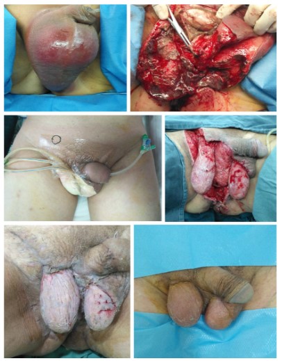
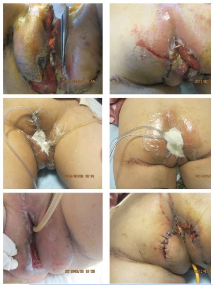
Typical cases
There were three typical cases: two male patients, including
one with a perianal abscess (Figure 1, patient #3) and one with
perineal injury (Figure 2, patient #21), and one female patient
with perineal surgery (Figure 3, patient #5).
Discussion
This retrospective study aimed to analyze FG's diagnosis and
treatment for the first diagnosis and referral at a tertiary hospital.
The results revealed that FG is a rare but severe necrotizing soft
tissue infection. Early accurate diagnosis and early debridement
surgery are necessary to reduce hospitalization time, course of
disease, complications, and mortality.
Necrotizing soft tissue infections are a group of diseases related to necrotizing changes in any layer of soft tissue lumen, including simple skin necrosis and life-threatening fascia and muscle
infections [1-3]. It features acute onset, rapid progression, and
high mortality. Necrotizing fasciitis is a severe infectious condition, mainly involving superficial fascia and subcutaneous tissue [1-3]. Necrotizing fasciitis that occurs in the perineal, genital, and
perianal regions are often called FG. The disease is characterized
by rapid progress and explosive infection, accompanied by sepsis.
If the diagnosis is delayed and the operation is not carried out in
time, the mortality rate will be high. Literature reports that 40%
of patients have sepsis, and the mortality rate ranges from 20% to
70%-80% [11-16]. In the present study, there were cases of respiratory failure. After a period of anti-infection treatment, the local
abscess was broken. The local infection was controlled, and the
whole body condition deteriorated in three patients, ultimately
leading to death. Nevertheless, the mortality rate was 14%, which
is lower than the reported 15% to 70%-80% [11-16,29-32]. Of
note, Kuzaka et al. 33 reported no deaths among their 13 patients. This could be due to an early diagnosis and treatments in
a tertiary hospital. Four patients were referred from primary and
secondary hospitals. Still, we cannot exclude the possibility that
some patients were not recognized in time in other hospitals and
that the patients died before being referred to our tertiary hospital. Many pathogenic bacteria are often isolated and cultured locally in FG lesions, cooperating to destroy the tissues and secrete
various toxins and metabolites, resulting in occlusive endarteritis
and skin and subcutaneous vascular thrombosis, causing necrosis.
Tissue necrosis and the infection spread along the fascia plane,
initially involving the superficial (Colles fascia) and deep fascia of
the genitals [34]. Later, it spreads to the covered skin and even
involves muscles. Colles fascia infection can spread to the penis
and scrotum through Buck’s and Dartos fascia or to the anterior abdominal wall through Scarpa’s fascia [34]. Because of the different blood supply sources and fascia levels of the scrotum,
penile skin, testis, and corpus cavernosum, the testis and corpus
cavernosum are less involved. Once testis is infected, it indicates
retroperitoneal origin or transmission of infection [35-37]. In the
present study, orchiectomy had to be performed in two patients.
There are many sources of FG, such as anorectal, genitourinary,
or gynecology. The most common anorectal source is the perianal abscess. Genitourinary factors include an indwelling catheter, traumatic catheter insertion, long-term urethral stricture,
perineal trauma, and human bite or scratch. Female FG patients
often originate from infected Babbitt glands, septic abortion,
perineal incision wound, sexual intercourse injury, or genital resection. Inducing factors are mainly patients’ comorbidities such
as diabetes, malnutrition, medical immunosuppression (such as
chemotherapy, long-term use of steroids, malignant tumors), HIV
infection, leukemia, liver diseases, and uremia [12,14,17]. A study
from Chinese Taiwan reported that the most common source of
infection is the gastrointestinal tract (30%-50%), followed by the
genitourinary tract (20%-40%), and skin injury (20%). The morbidity proportion of males to females was 10:1 [13]. In this study,
21/22 patients were males, as supported by the literature. The
exact source of infection was unfortunately unclear in many patient charts and could not be analyzed. Early and correct diagnosis includes complete laboratory tests, imaging examination, and
bacteriological examination, such as blood routine, coagulation
spectrum, CRP, PCT, blood sugar, and renal function. The examination includes package expansion ultrasound, plain film, Computed
Tomography (CT), and Magnetic Resonance Imaging (MRI). A plain
film can show gas in soft tissue [2,4,5,18]. CT may show fascia air
or gas, soft-tissue edema, or enhancement of fascia. CT and MRI
are time-consuming and can easily delay surgery. CT is associated
with a sensitivity of 94.3% (95% CI 81.2%-98.5%) and a specificity of 76.6% (95% CI 21.3%-97.5%) for the diagnosis of necrotizing fasciitis [38]. Point of care ultrasound (POCUS) is a relatively
new bedside diagnosis method, which can even find deep image
changes that cannot be captured by CT and MRI [39,40]. It should
be investigated in future studies Early correct diagnosis, active
surgical expansion, the correct treatment of inducing factors and
comorbidities, combined application of broad-spectrum antibiotics, and comprehensive treatment of severe sepsis are the keys to
successful treatment [2,4,5,18]. If blisters, hypotension, and other systemic changes occur within a few hours, surgery should be
performed as soon as possible under adequate fluid resuscitation
[2,4,5,18]. If the progression is not obvious, a tissue biopsy should
be performed. During wound exploration, tissue integrity and infiltration depth need to be evaluated. Necrotic skin and subcutaneous fascia tissue of the scrotum, penis, and perineum need to
be removed, leading to a severe loss of skin and soft tissue and
require reconstruction surgery. Some authors reported that an
average of 3-3.5 operations is required per patient [10,37]. Still,
only 1.6 was performed in the present study, which could be due
to the intervention's higher initial aggressiveness. Early diagnosis
and aggressive treatment are advocated by Yucel et al. [41] and
Heijkoop et al. [42]. Koukouras et al. reported that the colostomy
rate was 55.5%, the cystostomy ratewas 37.7%, and the orchiectomy rate was 26.6% [43]; the rates of such operations were lower in the present study, possibly because of early diagnosis and
intervention.
NPWT is an active way to help wound repair. Nevertheless, although there was no significant difference in survival rate between
NPWT and non-NPWT, the application significance of NPWT cannot be discussed because one of the three patients who died underwent local incision and drainage, one did not have surgery, one
had local ulceration before admission, and there was no complete
debridement surgery. Nevertheless, Iacovelli et al. [44] indicated
that the use of NPWT had advantages in terms of wound closure
and overall survival. Additional studies are necessary to examine
this point. This study has limitations. It was a single-center retrospective study in a small number of patients. The data that could
be analyzed was limited to those in the charts. Multicenter studies are necessary.
Conclusion
FG is a rare and life-threatening necrotizing infection that requires early diagnosis to reduce morbidity and mortality. A high
degree of clinical suspicion, combined with anatomical knowledge, risk factors, and etiology, is necessary for an accurate diagnosis and management. Although FG is still a clinical diagnosis based on medical history and physical examination results,
relevant laboratory, and radiological investigations can be used
as useful auxiliary means. FG treatment is based on emergency
surgical consultation for debridement of necrotic tissue, broad-spectrum antibiotics, intravenous infusion when necessary, and
hemodynamic resuscitation of vasoactive drugs.
Declarations
Competing interests: All authors declare that they have no any
conflict of interests.
Funding: This study was supported by the Project of the Education Department of Zhejiang Province (No. Y201431351), and the
Project of the Science and Technology Department of Zhejiang
Province (No. LGF20H190004).
Acknowledgements: None.
References
- Andrews L, Arora S. Images in emergency medicine. Male with
throat pain and neck swelling. Necrotizing fasciitis in association
with Ludwig’s angina. Ann Emerg Med. 2015; 65: e5-6.
- Stevens DL, Bisno AL, Chambers HF. Practice guidelines for the diagnosis and management of skin and soft tissue infections: 2014
update by the infectious diseases society of America. Clin Infect
Dis. 2014; 59: 147-159.
- Morgan MS. Diagnosis and management of necrotising fasciitis: a
multiparametric approach. J Hosp Infect. 2010; 75: 249-257.
- Bonne SL, Kadri SS. Evaluation and Management of Necrotizing
Soft Tissue Infections. Infect Dis Clin North Am. 2017; 31: 497-511.
- Peetermans M, de Prost N, Eckmann C, Norrby-Teglund A, Skrede
S, et al. Necrotizing skin and soft-tissue infections in the intensive
care unit. Clin Microbiol Infect. 2020; 26: 8-17.
- Ersoz F, Sari S, Arikan S, Altiok M, Bektas M, et al. Factors affecting mortality in Fournier’s gangrene: experience with fifty-two patients. Singapore Med J. 2012; 53: 537-540.
- Aridogan IA, Izol V, Abat D, Karsli O, Bayazit Y, et al. Epidemiological
characteristics of Fournier’s gangrene: a report of 71 patients. Urol
Int. 2012; 89: 457-461.
- Bednarek M, Drozdz W. [A rare case of the extensive Fournier’s
gangrene developed in the course of a perianal abscess]. Przegl
Lek. 2008; 65: 410-412.
- Mazur A, Karbowski M. [Fournier’s gangrene-a case report]. Wiad
Lek. 2013; 66: 260-261.
- Sroczynski M, Sebastian M, Rudnicki J, Sebastian A, Agrawal AK. A
complex approach to the treatment of Fournier’s gangrene. Adv
Clin Exp Med. 2013; 22: 131-135.
- Sabzi Sarvestani A, Zamiri M, Sabouri M. Prognostic Factors for
Fournier’s 14 Gangrene; A 10-year Experience in Southeastern
Iran. Bull Emerg Trauma. 2013; 1: 116-122
- Thwaini A, Khan A, Malik A, Cherian J, Barua J, et al. Fournier’s
gangrene and its emergency management. Postgrad Med J. 2006;
82: 516-519.
- Chen SY, Fu JP, Wang CH, Lee TP, Chen SG. Fournier gangrene: a
review of 41 patients and strategies for reconstruction. Ann Plast
Surg. 2010; 64: 765-769.
- Mallikarjuna MN, Vijayakumar A, Patil VS, Shivswamy BS. Fournier’s Gangrene: Current Practices. ISRN Surg. 2012; 2012: 942437.
- Stephens BJ, Lathrop JC, Rice WT, Gruenberg JC. Fournier’s gangrene: historic (1764-1978) versus contemporary (1979-1988) differences in etiology and clinical importance. Am Surg. 1993; 59:
149-154.
- Garcia Marin A, Turegano Fuentes F, Cuadrado Ayuso M, et al. Predictive factors for mortality in Fournier’ gangrene: a series of 59
cases. Cir Esp. 2015; 93: 12-17
- Yanar H, Taviloglu K, Ertekin C. Fournier’s gangrene: risk factors
and strategies for management. World J Surg. 2006; 30: 1750-
1754.
- Sugihara T, Yasunaga H, Horiguchi H. Impact of surgical intervention timing on the case fatality rate for Fournier’s gangrene: an
analysis of 379 cases. BJUInt. 2012; 110: E1096-1100.
- Zhao JC, Zhang BR, Shi K, Zhang X, Xie CH, et al. Necrotizing soft
tissue infection: clinical characteristics and outcomes at a reconstructive center in Jilin Province. BMC Infect Dis. 2017; 17: 792.
- Baharestani MM. Negative pressure wound therapy in the adjunctive management of necrotizing fascitis: examining clinical outcomes. Ostomy Wound Manage. 2008; 54: 44-50.
- El-Sabbagh AH. Negative pressure wound therapy: An update.
Chin J Traumatol. 2017; 20: 103-107.
- Paula FM, Pinheiro EA, Oliveira VM, Ferreira CM, Monreal MTFD,
et al. A case report of successful treatment of necrotizing fasciitis using negative pressure wound therapy. Medicine (Baltimore).
2019; 98: e13283.
- Novelli G, Catanzaro S, Canzi G, Sozzi D, Bozzetti A. Vacuum assisted closure therapy in the management of cervico-facial necrotizing fasciitis: a case report and review of the literature. Minerva
Stomatol. 2014; 63: 135-144.
- Steinstraesser L, Sand M, Steinau HU. Giant VAC in a patient with
extensive necrotizing fasciitis. Int J Low Extrem Wounds. 2009; 8:
28-30.
- Argenta LC, Morykwas MJ. Vacuum-assisted closure: a new method for wound control and treatment: clinical experience. Ann Plast
Surg 1997; 38: 563-576.
- Lima R, Coltro PS, Farina JAJ. Negative pressure therapy for the
treatment of complex wounds. Rev Col Bras Cir. 2017; 44: 81-93.
- Czymek R, Schmidt A, Eckmann C, et al. Fournier’s gangrene: vacuum-assisted closure versus conventional dressings. Am J Surg.
2009; 197: 168-176.
- Huang C, Leavitt T, Bayer LR, Orgill DP. Effect of negative pressure
wound therapy on wound healing. Curr Probl Surg. 2014; 51: 301-
331.
- Wetterauer C, Ebbing J, Halla A, et al. A contemporary case series
of Fournier’s gangrene at a Swiss tertiary care center-can scoring systems accurately predict mortality and morbidity? World J
Emerg Surg. 2018; 13: 25.
- Wang L, Han X, Liu M, et al. Experience in management of Fournier’s gangrene: a report of 24 cases. J Huazhong Univ Sci Technolog
Med Sci. 2012; 32: 719-723.
- Benjelloun el B, Souiki T, Yakla N, et al. Fournier’s gangrene: our
experience with 50 patients and analysis of factors affecting mortality. World J Emerg Surg. 2013; 8: 13.
- Madsen MB, Skrede S, Perner A, et al. Patient’s characteristics and
outcomes in necrotising soft-tissue infections: results from a Scandinavian, multicentre, prospective cohort study. Intensive Care
Med. 2019; 45: 1241-1251.
- Kuzaka B, Wroblewska MM, Borkowski T. Fournier’s Gangrene:
Clinical Presentation of 13 Cases. Med Sci Monit. 2018; 24: 548-
555
- Katib A, Al-Adawi M, Dakkak B, Bakhsh A. A three-year review of
the management of Fournier’s gangrene presented in a single Saudi Arabian institute. Cent European J Urol. 2013; 66: 331-334.
- Eke N. Fournier’s gangrene: a review of 1726 cases. Br J Surg. 2000;
87: 718-728.
- Eke N, Echem RC, Elenwo SN. Fournier’s gangrene in Nigeria: a review of 21 consecutive patients. Int Surg. 2000; 85: 77-81.
- Chawla SN, Gallop C, Mydlo JH. Fournier’s gangrene: an analysis of
repeated surgical debridement. Eur Urol. 2003; 43: 572-575.
- Fernando SM, Tran A, Cheng W, et al. Necrotizing Soft Tissue Infection: Diagnostic Accuracy of Physical Examination, Imaging, and
LRINEC Score: A Systematic Review and Meta-Analysis. Ann Surg.
2019; 269: 58-65.
- Kehrl T. Point-of-care ultrasound diagnosis of necrotizing fasciitis
missed by computed tomography and magnetic resonance imaging. J Emerg Med. 2014; 47: 172-175.
- Magalhaes L, Martins SRP, Nogue R. The role of point-of-care ultrasound in the diagnosis and management of necrotizing soft tissue
infections. Ultrasound J. 2020; 12: 3.
- Yucel M, Ozpek A, Basak F, et al. Fournier’s gangrene: A retrospective analysis of 25 patients. Ulus Travma Acil Cerrahi Derg. 2017;
23: 400-404.
- Heijkoop B, Parker N, Spernat D. Fournier’s gangrene: not as lethal
as previously thought? A case series. ANZ J Surg. 2019; 89: 350-
352.
- Koukouras D, Kallidonis P, Panagopoulos C, et al. Fournier’s gangrene, a urologic and surgical emergency: presentation of a multi-institutional experience with 45 cases. Urol Int. 2011; 86: 167-172.
- Iacovelli V, Cipriani C, Sandri M, Filippone R, Ferracci A, et al. The
role of vacuum-assisted closure (VAC) therapy in the management
of FOURNIER’S gangrene: a retrospective multi-institutional cohort study. World J Urol. 2020.