Introduction
Burns can be classified according to the depth of the damage
caused to the skin and underlying tissues.
Second degree burns heal by re-epithelialization, at the edges
of the wound the basal cells begin to migrate towards the viable
wound bed, they are stimulated by the loss of cell contact inhibition, by the release of local growth factors (factor growth factor, transforming growth factor alpha), and contact with proteins
from the wound bed, among others. However, the migration limit
from the edges of the wound is 1-2 cm. When the cutaneous appendages remain viable, keratinocytes can migrate from them to
the wound, likewise keratinocytes migrate more quickly in a bed
that is maintained with adequate moisture [1].
Another way to classify burns is according to their etiology:
heat, electrical, and chemical. Electrical burns are rare, corresponding to less than 5% of all burns, they represent the most
frequent cause of amputation in burn patient management
units. They are classified as high voltage (>1,000 V) or low voltage (<1,000 V) injuries. Those with high voltage present more extensive damage, injuring deeper soft tissues, therefore they will
require a greater number of surgical interventions [2]. An electric
arc can reach temperatures of up to 2,000°C, causing thermal and
electrical burns that can vary in depth and cause damage to internal organs [3]. The resistance of the tissues in ascending order
is: nerves, blood vessels, muscle, skin, tendons, fat, bone. There
are three mechanisms that cause electrical burns: a true electrical injury caused by the flow of current against the resistance of
the involved tissues causing damage due to excessive heating of
these, a direct thermal burn due to the heat of the arc that occurs
when the high voltage current passes through the air, and lastly
the thermal burn produced by the igniting clothing of the patient
or ignited surroundings [4].
For the treatment of burn injuries there are dressings, dermal
analogues, temporary skin substitutes; which favor the re-epithelialization of the wound bed, accelerating the healing process and
some of them having specific properties.
The human amniotic membrane is a temporary skin substitute
that has low antigenicity, high antimicrobial potential, has antiinflammatory properties that reduce fibrosis, likewise isolates the
injured bed from the external environment, reduces heat and fluid loss, and favors the epithelialization of lesions from previously
excised 2nd and 3rd degree burn wounds. It is obtained from the
delivery rooms and later undergoes a process of radio sterilization
and cryopreservation in the tissue bank [5].
The fetal human amniotic membrane is composed of 2 parts:
The chorion and the amnion. The chorion, which is the external
layer providing a sac-like appearance, composed of trophoblastic and mesenchymal tissue and the amniotic membrane corresponding to the internal layer. The amnion is composed of 5 layers: epithelium, basement membrane and stromal matrix, the latter is divided into a compact, fibroblastic and spongy layer [6,7].
The epithelium performs 3 main functions: covering, secretory
activity, intense intercellular and transcellular transport. It produces growth factors such as beta transforming growth factor, hepatocyte growth factor, platelet-derived growth factor, epidermal
growth factor, keratinocyte growth factor, among others.
The basement membrane facilitates epithelial cell migration,
promotes epithelial differentiation, prevents epithelial apoptosis
and injury site expansion, and decreases pain [8].
The stromal matrix is avascular, producing growth factors such
as epidermal growth factor, hepatocyte growth factor, and keratinocyte growth factor. Suppresses inflammatory cells by rapid
stimulation of apoptosis, contains various forms of protease inhibitors; reducing granulation tissue and excessive angiogenesis,
thus decreasing the formation of fibrosis, which would manifest
as hypertrophic pathological scarring in the burned patient.
The amnionic membrane does not integrate with the lesion
bed and does not vascularize, but it favors angiogenesis and induces the formation of granulation tissue (both in a controlled
manner) and epithelialization due to the presence of multiple
growth factors: Epidermal Growth Factor (EGF), Transforming
Factor Alpha (TGF-α), Transforming Factor Beta 1 (TGF-β1), Keratinocyte Epidermal Growth Factor Receptor (KGFR), Hepatocyte
Epidermal Growth Factor Receptor (HGFR) [9].
It has a very low risk of rejection, due to its low antigenicity
and lack of HLA-A, HLA-B, HLA-C and HLA-DR. It decreases the local inflammatory response at its site of application by inducing
inflammatory cell apoptosis, and the phenotypic change of macrophages from a pro-inflammatory M1 type to an anti-inflammatory/regulatory M2 phenotype.
It also has the property of acting as an insulator, since it protects and preserves a clean excised wound, it has also been observed to decrease local pain [10].
Case presentation
A 41-year-old male patient who suffered a high-voltage electrical burn. His condition began when he was working, the truck in
which he was traveling was trapped between high-voltage cables,
when maneuvering to try to free the vehicle, he presented an
electric shock from the high-voltage cable and was ejected from
the vehicle at a distance of approximately 3 meters. He is assisted
by paramedical personnel and transferred by ambulance helicopter to ISSEMyM Toluca Medical Center, our center being the 3rd
level reference hospital in the State of México.
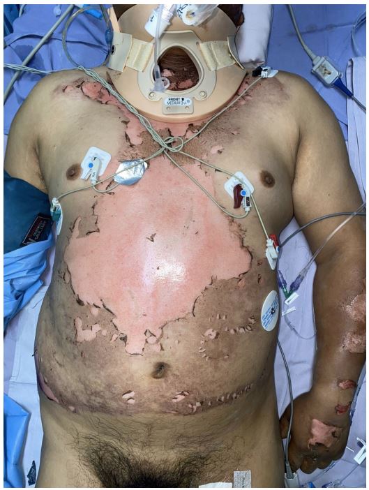
During his admission to the shock area of the emergency department, he presented mixed superficial and deep 2nd degree
burns, 3rd degree of 19% of the total body surface burned: face
1%, neck 1%, thorax and anterior abdomen 9%, right upper limb
1%, left upper limb 1%, abdomen 2%, right lower limb 0.5%, circumferential left lower limb 3.5%. Burned vibrissae, peri-oral and
palate burns were observed, these being signs indicative of airway
burn, for which orotracheal intubation was decided (Figure 1).
He underwent 15 days of hospital stay in charge of the Reconstructive Surgery service, requiring 9 days of management in the
of which 6 days he underwent invasive mechanical ventilation. On
the day of his admission, wound washing was performed in the
intensive care unit bed because the patient was hemodynamically
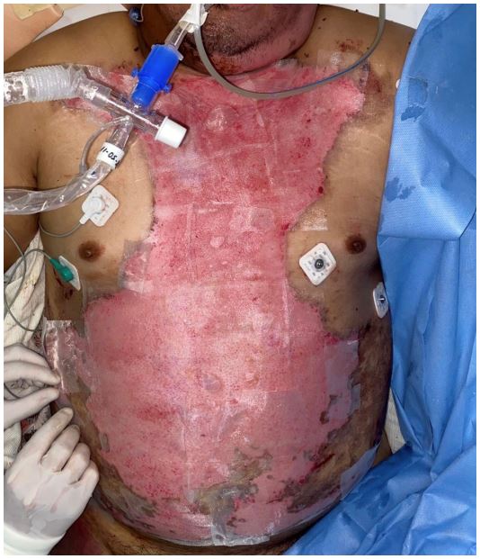
unstable. 1 day after his admission, a forearm and right hand fasciotomy was performed, 2 days later fasciotomy closure was performed; 7 days after his admission, surgical cleaning and amnion
placement were performed on burns in the thorax and anterior
abdomen, without being removed (Figure 2); with clinical improvement: without presenting data of local or systemic infection,
notable decrease in pain and adequate adherence to the affected
area and presence of underlying granulation tissue.
Currently, 10 months after the electrical burn, the patient
presents complete recovery from the burns, hyperchromic scars,
no skin retraction, no raised edges, and no hypertrophic scarring
(Figure 3). Presents neurological sequelae consisting of polyneuropathy secondary to electrocution, which are managed by the
neurology service. It is also found in rehabilitation therapies to
improve the gait pattern, increase strength, and improve ranges
of motion in the upper and lower extremities.
Discussion
Regarding our clinical case presented, the human amniotic
membrane is used as a temporary skin substitute in the treatment of patients with extensive burn injuries in general. To mention some of the main advantages of amniotic membrane in the
management of burns: it shortens the patient's recovery time, it
prevents fluid and electrolyte disturbances, it decreases the intensity of pain, it is almost completely transparent, which allows
us to observe the underlying wounds, it is very flexible and adapts
to the irregularities of the body surface (including joints), it is not
necessary to remove it [11,12]. It can also be used in a complementary way on a meshed skin autograft, to favor the healing of
the burned bed and improve the quality of healing [13].
It is easily accessible, the cost is minor, currently due to its bactericidal qualities and tissue bank protocols, the prevalence of infections has decreased.
Likewise, the results observed in the healing characteristics
have been favorable with respect to the almost null presence of
pathological scars, this is mainly due to the fact that the TGF-β1
released by the amniotic membrane inhibits the differentiation of
myofibroblasts into fibroblasts [14]. As we can see in our patient, who despite having presented second and third degree burns, did
not present hypertrophic scarring, the final result of the healing
is adequate, he only presented a slight hyperpigmentation of the
scars, being a favorable cosmetic result.
Conclusion
The human amniotic membrane is a very useful skin substitute
with great advantages in the management of patients with burns,
reducing their hospital stay by shortening the re-epithelialization
time of burned areas and preventing bed infection, reducing patient pain. Since it does not have to be removed, the need to use
skin grafts decreases, thus reducing morbidity since skin donor
areas will not be required.
Likewise, the results observed in the healing characteristics
have been favorable with respect to the almost null presence of
pathological scars, with hypertrophic scarring secondary to second and third degree burns being frequent.
References
- Greenhalgh, D. Management of Burns. N Engl J Med. 2019; 380: 2349-2359.
- Shih JG, Shahrokhi S, Jeschke MG. Review of adult electrical burn injury outcomes worldwide: An analysis of low-voltage vs highvoltage electrical injury. J Burn Care Res. 2017; 38: 293-298.
- Pielesz A, Gawłowski A, Biniaś D, Bobiński R, Kawecki M, et al. A Histologic Perspective on Electrical and Thermal Burn-Injured Human Skin. Adv Skin Wound Care. 2019; 32: 1-7.
- Arnoldo BD, Purdue GF. The diagnosis and management of electrical injuries. Hand Clin. 2009; 25: 469-79.
- Sandoval J, Ortega S, Balmelli B. Uso de membrana amniótica como cobertura temporal en pacientes pediátricos con quemaduras. An. Fac. Cienc. Méd. (Asunción), Agosto. 2022; 55: 59-67
- Alsina-Gibert M, Pedregosa-Fauste S. Aplicación de membrana amniótica en el tratamiento de las úlceras crónicas de extremidades inferiores. Actas Dermo-Sifiliográficas. 2012; 103: 608-613.
- Castellanos G, Bernabé-García N, Moraleda JM, Nicolás FJ. Amniotic membrane application for the healing of chronic wounds and ulcers. Placenta. 2017; 59: 146-153.
- Zelen C, Serena T, Setterolf D. Dehydrated human amnion/chorion membrane allografts in patients with chronic diabetic foot ulcers: A long-term follow-up studym. Wound Medicine. 2014; 4: 1-4.
- Fairbairn N, Randolph M, Redmond R. The clinical applications of human amnion in plastic surgery. Journal of Plastic, Reconstructive & Aesthetic Surgery. 2014; 67: 662-675.
- Gaviria-Castellanos JL, Gómez-Ortega V, Guerrero-Serrano L. Manejo de quemaduras faciales de segundo grado con membrana amniótica preservada en glicerol 85%. 2018; 44: 401-8
- Eskandarlou M, Azimi M, Rabiee S, Seif Rabiee MA. The Healing Effect of Amniotic Membrane in Burn Patients. World J Plast Surg. 2016; 5: 39-44.
- Mohammadi AA, Seyed Jafari SM, Kiasat M, Tavakkolian AR, Imani MT, et al. Effect of fresh human amniotic membrane dressing on graft take in patients with chronic burn wounds compared with conventional methods. Burns. 2013; 39: 349-353.
- Yang C, Xiong AB, He XC, Ding XB, Tian XL, et al. Efficacy and feasibility of amniotic membrane for the treatment of burn wounds: A meta-analysis. J Trauma Acute Care Surg. 2021; 90: 744-755.
- Muñoz-Torres JR, Martínez-González SB, Lozano-Luján AD, Martínez-Vázquez MC, Velasco-Elizondo P, et al. Biological properties and surgical applications of the human amniotic membrane. Front Bioeng Biotechnol. 2023; 10: 1067480.